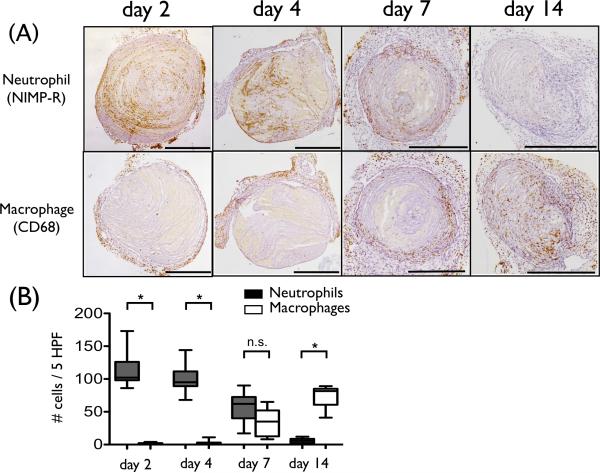
Figure 4.
Recruitment of inflammatory cells into DVT. (A) Representative immunostaining of neutrophil and macrophage from various DVT timepoints. Neutrophils are abundant and predominate in early day 2-4 DVT. Thrombus macrophages were evident from day 7 and resided at the outer DVT edge. (B) The number of neutrophils (black) and macrophages (white) per 5 HPF (high power field) were shown. (*p<0.0001). Scale bar, 500 μm. n.s.=not significant.

