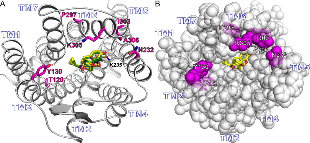
Figure 7. The relationship of the naltrexone with the mutated residues.
A) Rendering of the murine μ opioid receptor. The seven residues that were mutated back to their native residue identities are in magenta (E120T2.54, K130Y2.64, D232N5.36, E297P6.50, K303I6.56, G305K6.58, and K306A6.59). The docking pose of naltrexone (yellow) overlaps with the pose of β-Funaltrexamine (green). B) Similar view as (A) but with the protein rendered in space filling representation. The docked posed of naltrexone (yellow) does not directly contact any of the seven mutated positions (magenta).

