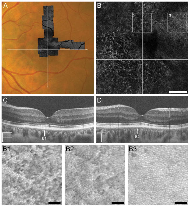
Figure 2.
Case 2, TC_10006. Trauma-induced diffuse disruption of the EZ and IZ bands and ONL thinning throughout the macula and on SD-OCT corresponds to the level of photoreceptor disruption on AOSLO 5 months after cgBOT. A. Full extent of AOSLO montage overlaid onto a color fundus photograph of the left eye with white arrows indicating the position of SD-OCT line scans (C, D). Central portion of AOSLO montage (B) showing widespread disruption of the foveal and perifoveal photoreceptor mosaic. Numbered white boxes indicating the position of the AOSLO insets (B1 to B3), and white lines again indicate the position of SD-OCT line scans (C, D). Scale bar = 200 μm. AOSLO insets, B1 and B2, taken from ~1–1.5° eccentricity show a disrupted cone mosaic of reduced density (18,281 cones/mm2 and 17,812 cones/mm2, respectively). Normal cone density for this location is 43,582 ± 6,521cones/mm2.23 The hyporeflective cones correspond to a region of IZ band disruption on SD-OCT (arrows B1 and B2 in panels C and D, respectively). AOSLO inset B3 shows a perifoveal (~2° eccentricity) contiguous cone mosaic of normal density (27,500 cones/mm2). Normal cone density for this location is 25,721 ± 3,506 cones/mm2.23 Although not captured with the SD-OCT line scans shown here, B3 corresponds to normal outer retinal lamination seen on volumetric SD-OCT through the macula. Scale bars in AOSLO insets B1–B3 = 40 μm.

