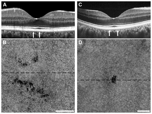
Figure 7.
Subclinical photoreceptor defects following cgBOT. Subjects RR_0045, Case 8 (A, B) and SH_0874, Case 9 (C, D) had visual complaints despite normal clinical imaging with SD-OCT. Subject RR_0045 had a normal-appearing SD-OCT acquired using Spectralis (A). Subject SH_0874 had normal-appearing SD-OCT on both Spectralis and Cirrus, while imaging using the Bioptigen SD-OCT revealed a small focal disruption of the EZ and IZ (C). Vertical arrows indicate the area subtended by the AOSLO montages (B, D). The AOSLO images are displayed on a logarithmic scale and each shows clear photoreceptor disruption. Subject RR_0045 (B) had two areas of diffuse photoreceptor hyporeflectivity, while subject SH_0874 (D) had a small discrete lesion. The dashed black line on each AOSLO image indicates the location of the corresponding SD-OCT image. Scale bars = 100 μm.

