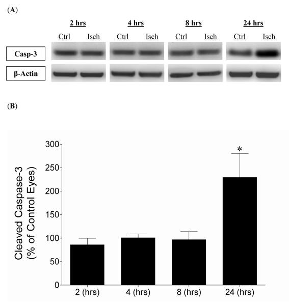
Figure 3.
Effect of 45 minutes of acute ischemia on retinal cleaved caspase-3 levels 2, 4, 8, and 24 hours from ischemia initiation. (A) Representative Western blot of retinal lysates for cleaved caspase-3 and β-actin at 2, 4, 8, and 24 hours after initiation of ischemic injury. (B) Levels are expressed as a mean percentage of the ischemic eyes relative to the control eyes with no significant changes at 2 hours (85.9 ± 13.6; n=4), 4 hours (100.8 ± 8.2; n=5), and 8 hours (96.8 ± 17.2; n=7). Significant increases (* P <0.05) in cleaved caspase-3 occurred at 24 hours (229.4 ± 51.1; n=9) after initiation of ischemia (Student t-test). Abbreviations: Cleaved caspase-3 (Casp-3); Control eyes (Ctrl); Hours post-ischemia initiation (hrs); Ischemic eyes (Isch).

