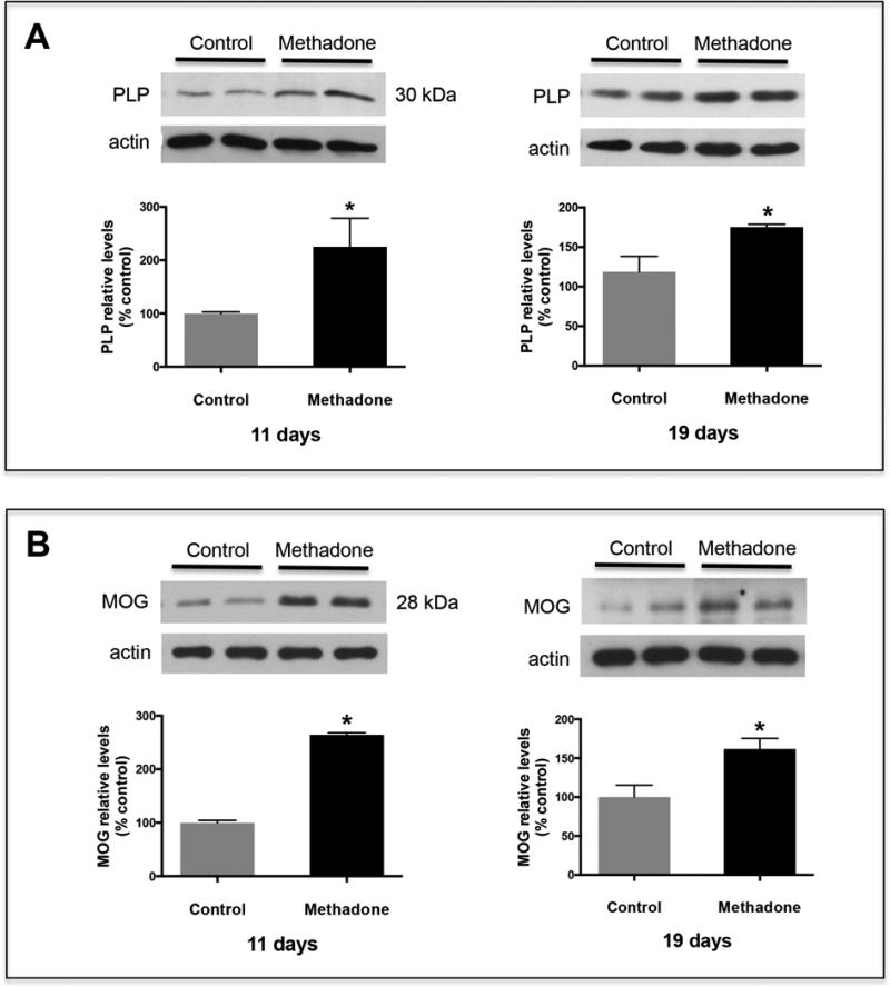
Figure 2. Pups exposed to methadone also exhibit increased brain levels of PLP and MOG.
Pups were perinatally exposed to methadone as described under “Methods”. Total brain homogenates from 11 and 19-day-old rats were analyzed for PLP (A) and MOG (B) expression by western blot analysis with the respective antibodies. The figures show representative western blots. The bar graphs depict the results as % of those corresponding to the control animals. Each value corresponds to the mean ± SEM from at least 10 brains from 3 different litters/group. β-actin levels were used as loading controls. Control vs. methadone, p < 0.03.

