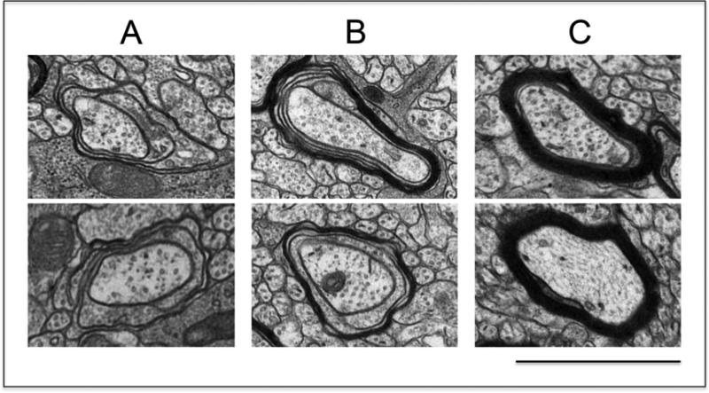
Figure 3. Classification of axons according to their stage of myelination.
Brain samples for analysis of myelinated axons were prepared from 16-day-old pups and thin sections of the region containing the mid body of the corpus callosum were examined by electron microscopy as described in the “Methods” section. Samples were photographed at 5,000 X magnification and axons were classified within the following three categories: (A) axons at the initial stage of myelination, as represented by the presence of no more than four loose wraps of membrane; (B) axons at intermediate stages of myelin formation, as indicated by the presence of more than four but still uncompacted membrane wraps; and (C) axons with already compacted myelin sheaths. (bar = 1μm). The photos were taken from samples corresponding to the control animals.

