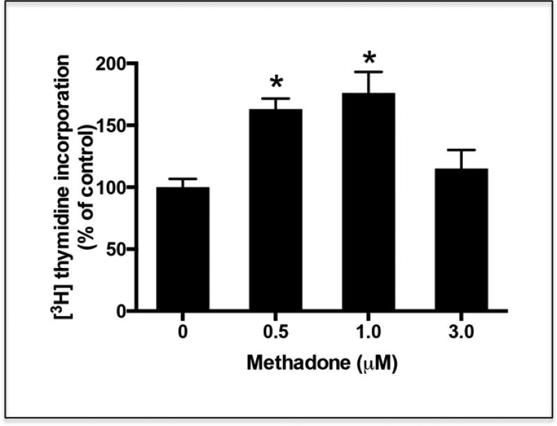
Figure 5. Treatment of oligodendrocyte progenitors with methadone results in increased cell proliferation.
Oligodendrocyte progenitors were isolated from 3-day-old rat brain as indicated under “Methods”. One day after isolation, the cells were incubated for 24 hr in CDM supplemented with [3H]thymidine in the presence or absence of different concentrations of methadone. Cell proliferation was evaluated by measuring [3H]thymidine incorporation into the DNA. The results are expressed as percentage of the control values ± SEM from at least 3 different experiments. *p<0.005. Control vs. 3.0 μM, N.S.

