
FIGURE 7.
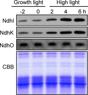
Amounts of NdhI, NdhK, and NdhO in WT cells grown under different irradiances. The protein levels of NdhI, NdhK, and NdhO were analyzed by Western blot on WT cells grown under growth light (40 μmol photons m−2 s−1) before (2 and 0 h) and after transfer to high light (200 μmol photons m−2 s−1) for 2, 4, and 6 h. Lanes were loaded with thylakoid membrane proteins corresponding to 1 μg of chlorophyll a. In the lower panel, a replicate gel stained with Coomassie Brilliant Blue (CBB) was used as a loading control.