FIGURE 3.
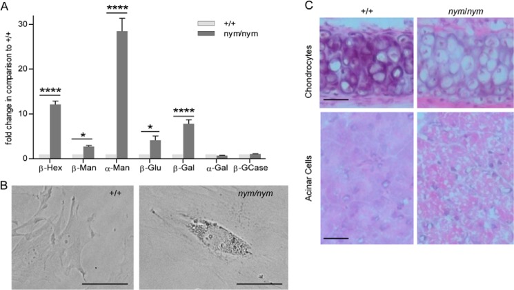
Increased lysosomal enzyme activity in blood sera and inclusion bodies present in mouse fibroblasts, secretory and connective tissue. A, the relative enzymatic activities of the lysosomal hydrolases β-Hexosaminidase (β-Hex), β-mannosidase (β-Man), α-mannosidase (α-Man), β-glucuronidase (β-Glu), β-galactosidase (β-Gal), α-galactosidase (α-Gal), and β-glucocerebrosidase (β-GCase) were measured in blood sera of 3-month-old wild-type (+/+) and nym (nym/nym) mice. The specific activities of the wild-type were set to 1. Values are expressed as mean ± S.E. (error bars) (n = 8; *, p < 0.05; **, p < 0.01; ***, p < 0.001; ****, p < 0.0001). B, light microscopy imaging of MEFs isolated from wild-type and nym embryos (E12.5). Accumulation of inclusion bodies present in the nym MEFs. Scale bar, 40 μm. C, top, the cytoplasm of hypertrophic chondrocytes is distended by microvacuoles in the nym mouse. These inclusions are aggregates of polysaccharides that increase in storage material with age. Bottom, marked disorganization of the pancreas in the nym mouse with tightly packed cells distended by large vacuoles. Scale bar, 50 μm.

