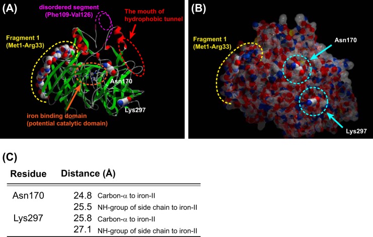
FIGURE 8.
Locations of Asn170 and Lys297 and the F1 fragment in an RPE65 three-dimensional structure model. The locations of two identified key residues and the F1 fragment are shown in the three-dimensional model based on the crystal structure of bovine RPE65 (Protein Data Bank entry 3FSN). The iron binding site, within the catalytic domain, is indicated by an orange dotted circle. The disordered segment (Phe109–Val126), which contains a palmitoylated Cys residue (Cys112), is shown by a pink dotted line. The entrance of the hydrophobic tunnel containing an active site is indicated by a red dotted circle. The location of the F1 fragment is indicated by the yellow dotted line. Both Asn170 and Lys297 residues are located on the surface of the protein (A and B) and more than 20 Å distant from iron(II) in the potential catalytic domain (C).

