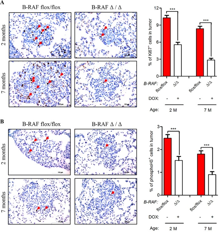
FIGURE 6.
Reduced proliferation in C-RAF BxB lung tumors of DOX-induced compound mice. A and B, SpC-C-RAF BXB/SpC- rtTA/Tet-O-cre/B-RAFflox/flox compound mice were DOX-induced for 2 and 7 months and compared with aged-matched controls. Representative pictures and quantification of Ki67 (A), phospho-H3 (B) staining (brown) of paraffin-embedded lung sections. Genotype and induction status are as indicated. Red arrows point to positively stained cells. Hematoxylin was used as a counterstain. A total of 75 tumors from five mice for each group were analyzed. Mean values are + S.E.; t test, ns, not significant, ***, p < 0.0005.

