
FIGURE 10.
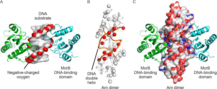
A proposed model of the Arn dimer·McrB DNA binding domain complex. A, the McrB DNA binding domain·DNA complex structure (PDB code 3SSC) contains two McrB DNA binding domains (shown in green and cyan) and a DNA substrate (shown as the surface). The complex structure revealed that the DNA-associating loops of two McrB DNA binding domains protrude into the DNA minor groove on two lateral parts of DNA. B, the β-carbons of Asp and Glu on Arn (showed as spheres) were manually aligned with the DNA-phosphate backbone (orange lines), and the DNA-matched β-carbons are highlighted in red. C, the Arn dimer in the Arn dimer·McrB DNA binding domain complex structure is shown in the surface mode and color-coded according to the atom type (white, carbon; red, oxygen; blue, nitrogen) to emphasize the three-dimensional space arrangement.