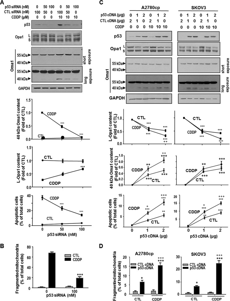
FIGURE 5.
p53-dependent action of CDDP on Oma1 (40 kDa) and L-Opa1 contents, mitochondrial fragmentation, and apoptosis in gynecologic cancer cells. A, chemosensitive CECA cells (OV2008) were transfected with p53 siRNA or control siRNA (CTL; scramble siRNA) (0–100 nm, 24 h) and subsequently cultured with CDDP (10 μm, 24 h) or control (DMSO). The contents of p53, Oma1, Opa1, and GAPDH (loading control) were examined by Western blotting. Apoptosis was examined by Hoechst assay. Knockdown of p53 inhibited CDDP-induced increase in Oma1 content (40 kDa; ***, p < 0.001) and loss in L-Opa1 content (*, p < 0.05; ***, p < 0.001; n = 3). p53 siRNA significantly decreased apoptosis induced by CDDP (***, p < 0.001, n = 3). More than 300 cells were assessed in each experimental group. B, OV2008 cells were treated with p53 siRNA or control siRNA as described in A and subsequently cultured with CDDP (10 μm, 6 h) or control (DMSO). Mitochondria were immunostained with Tom20 antibody. Mitochondrial phenotypes were examined by immunofluorescence confocal microscopy. Knockdown of p53 significantly decreased CDDP-induced mitochondrial fission (***, p < 0.001, n = 3). More than 100 cells were assessed in each experimental group. C, chemoresistant A2780cp (p53 mutant) and SKOV3 (p53 null) cells were transfected with WT-p53 cDNA or control cDNA (empty vector) (0–2 μg, 24 h) and subsequently treated with CDDP (10 μm, 24 h) or control (DMSO). Contents of p53, L-Opa1, and Oma1 were examined by Western blot. Apoptosis was examined by Hoechst assay. Reconstitution of WT-p53 not only induced Oma1 cleavage, L-Opa1 processing, and apoptosis in both CDDP-resistant OVCA cell lines (***, p < 0.001; *, p < 0.05, versus control plasmid, n = 3), but markedly sensitized them to CDDP-induced Oma1 cleavage, L-Opa1 processing, and apoptosis (+, p < 0.05; ++, p < 0.01, +++, p < 0.001, versus control). More than 300 cells were assessed in each experimental group). D, A2780cp (p53 mutant) and SKOV3 (p53 null) cells were transfected with WT-p53 cDNA or control cDNA (empty vector) (0.44 μg/well on 8-well chamber slide, 24 h) and subsequently treated with CDDP (10 μm, 6 h) or control (DMSO). Mitochondria were immunostained with Tom20 antibody and examined by immunofluorescence confocal microscopy. Reconstitution of WT-p53 not only increased percentage of cells with fragmented mitochondria in both CDDP-resistant OVCA cell lines (***, p < 0.001; *, p < 0.05, versus control plasmid, n = 3), but markedly sensitized them to CDDP-induced mitochondrial fragmentation (+++, p < 0.001, versus DMSO). More than 100 cells were assessed in each experimental group.

