FIGURE 1.
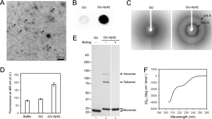
Characterization of GU-Aβ42 oligomers. A, transmission electron microscopy image of GU-Aβ42 oligomers shows globular structures with diameters of 10–12 nm. Arrows point to several of the oligomers. B, dot blot analysis with A11 antibody shows that GU-Aβ42 oligomers bind strongly to A11, while GU alone shows very weak reactivity. C, GU-Aβ42 oligomers show a powder x-ray diffraction pattern consistent with β-sheet structure, and GU alone shows very weak diffraction. D, GU-Aβ42 oligomers have weak binding to ThT. A.U., arbitrary unit. Error bars are standard deviations of three independent measurements. E, SDS-PAGE shows that, in addition to monomer, GU-Aβ42 oligomers contain SDS-resistant tetramer and hexamer. F, circular dichroism spectrum of the GU sample.

