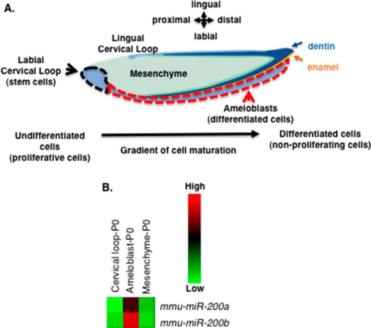
FIGURE 1.
miR-200a-3p expression is associated with differentiating dental epithelial cells. A, schematic of the mouse lower incisor cell and tissue structures. The black dotted line denotes the labial cervical loop (LaCL, stem cell niche), the red dotted line denotes the pre-secretory, secretory, and mature differentiated epithelial tissues (pre-ameloblasts and ameloblasts). Green shaded region, mesenchyme; dark blue, dentin; orange, enamel. B, heat map of selected miR-200a-3p and miR-200b-3p expression in the isolated dental epithelial tissue compartment (LaCL versus ameloblasts) and dental mesenchyme (ameloblast versus mesenchyme). These tissues were isolated from P0 mice lower incisors, total RNA was harvested, and miRs were analyzed by microRNA arrays. Five separate biological samples were analyzed. mmu, Mus musculus.

