
Figure 1.
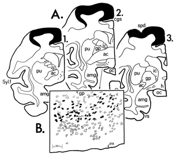
Three coronal sections from a rhesus monkey with HRP injected into Brodmann areas 4 and 6. A: The cortical areas within the injection site are blackened. Triangles represent nucleus basalis neurons. B: Camera lucida drawing from section 2 in A. Open profiles represent AChE-rich perikarya, blackened ones indicate double labeling of AChE-rich perikarya with retrogradely transported HRP. The original version of this figure was hand drawn in India ink by Gary Van Hoesen. ac, Anterior commissure; amg, amygdala; cgs, cingulate sulcus; gp, globus palidus; h, hypothalamus; ic, internal capsule; oc, optic chiasm; pu, putamen; rs, rhinal sulcus; spd, superior precentral dimple; Syl. f., Sylvian fissure. From Mesulam and Van Hoesen (1976) with permission.