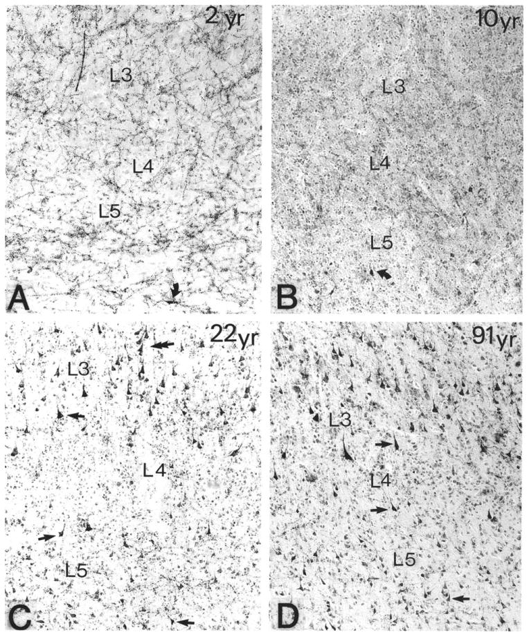
Figure 13.

Age-related differences in the density of AChE-rich neurons in the banks of the superior temporal sulcus in four human brains that came to postmortem examination at 2, 10, 22, and 91 years of age (A-D, respectively). The histochemical reaction was obtained with a modified Koelle-Friedenwald method, which provides better visualization of cell bodies than of axons. Straight arrows point to AChE-rich neurons. ×100. From Mesulam and Geula (1991) with permission.
