
Figure 14.
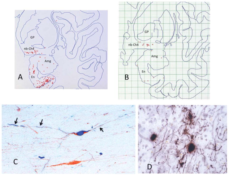
Early neurofibrillary degeneration of Ch4. A,B: Raw data obtained through the electronic plotter mapping of thioflavin-S stained whole-hemisphere sections in two autopsy cases aged 71 (A) and 72 (B). Neither had dementia or neurological disease. Each red cross marks a single NFT. These plots show that the nucleus basalis is at least as vulnerable to NFT formation as the entorhinal cortex and the amygdala. In A, there were no NFTs outside of the medial temporal lobe in that section. In B, the one additional NFT was encountered in the dorsal insula. C: Double immunostaining for ChAT (brown) and early neurofibrillary degeneration labeled with AT-8 (blue), an antibody that recognizes abnormally phosphorylated tau at early stages of neurofibrillary degeneration. The neuron on top is alive but undergoing neurofibrillary degeneration, whereas the one on the bottom is spared. The tauopathy has extended into the processes of the affected neuron (arrows). D: AChE histochemistry with a modified Karnovsky-Roots method in peristriate cortex of a 99-year-old woman who had no known dementia. Varicose cholinergic axons display dystrophic swollen profiles, which could represent early stages of cortical cholinergic degeneration and perhaps also aberrant attempts at regeneration. ×650. Amg, amygdala; En, entorhinal cortex; GP, globus pallidus; nb, nucleus basalis.