
Figure 15.
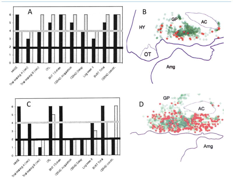
Cognitive correlates of tangles in the nucleus basalis. A and B are from a patient who died at the age of 95 years. A shows her test scores 3 years (black bars) and 4 months (gray bars) before death. On this graph, a level of 1 or 2 indicates performance that is impaired for age, levels 3 and 4 performance that is normal for age, and level 5 or 6 performance that is superior for age. In this patient, none of the scores was abnormal at either of the testing sessions. She was considered cognitively unimpaired. B, based on electronic plotting of stained tissue sections, shows normal neurons in her nucleus basalis (green circles) as well as those that contain NFT (red stars). C and D are from a patient who died at the age of 91 years. He was tested 2 years (black bars) and then 1 year (gray bars) before death. C shows that there has been a decline in performance and that he had multiple cognitive impairments (scores at level 2) at the last testing. Clinical notes indicate that the patient had progressed from a stage of mild cognitive impairment (MCI) at the first testing to early dementia of the AD type in the second. D shows his nucleus basalis at postmortem. Normal neurons are shown in green and neurons with NFT in red. Abbreviations for tests in frames A and C (from left to right): MMSE, Mini-Mental State Examination; Trail Making A and B, tests of executive function; CFL, test of verbal fluency; BNT, Boston naming test; CERAD Acquisition and Delay, tests of memory for words; Log Mem II, recall of a short story; BVRT, Benton visual retention test; CERAD constr., visuomotor test. AC, anterior commissure; Amg, amygdala; GP, globus pallidus; HY, hypothalamus; OT, optic tract. From Mesulam et al. (2004) with permission.