
Figure 16.
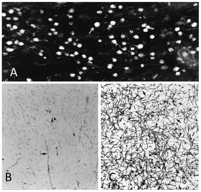
Neurofibrillary tangles and cholinergic innervation in AD. A: Nucleus basalis of a woman who died at the age of 84 years with clinical dementia and postmortem evidence of AD pathology. Histofluorescence in this thioflavin-S-stained section indicates that nearly all nucleus basalis neurons have been invaded by NFTs (arrow). Most are intracellular; others are ghost tangles. B: AChE histochemistry with a modified Karnovsky-Roots method shows nearly complete loss of cholinergic fibers in the middle temporal gyrus of the same patient. The arrow points to the one remaining axon in the field of view. C: Same region of the brain stained in the same fashion as in B but from a control subject who died at the age of 89 years with no evidence of dementia. The arrows point to two examples of cholinergic axons. ×100.