
Figure 7.
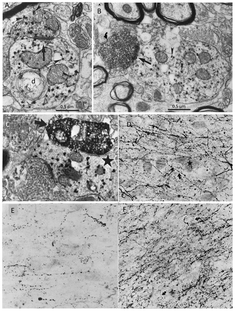
Transmitter-specific input into the monkey and human Ch4. A: ChAT immunoreactivity is visualized with the punctate VIP red reaction product (arrowheads) in the macaque monkey nucleus basalis. The ChAT axon on top is forming an asymmetric synapse (straight arrow) onto a postsynaptic ChAT dendrite (d) in the monkey Ch4. B: Double labeling of GAD (with diffuse DAB reaction product) and ChAT (with punctate VIP red reaction product) shows a GAD-positive GABAergic bouton (curved arrow) making a symmetric synapse (straight arrow) onto a nucleus basalis dendrite containing ChAT (arrowheads) in the macaque monkey brain. C: Double labeling of TH (with the diffuse DAB reaction product) and ChAT (with the punctate VIP red reaction product) showing a TH-immunoreactive bouton (arrow) forming a fine synapse onto a ChAT-immunoreactive nucleus basalis dendrite (star) in the macaque monkey. ×34,000. D: TH immunohistochemistry in the Ch4i sector of an autopsy specimen from a 27-year-old man showing multiple fine, varicose, TH-positive dopaminergic axons (curved arrow) coursing through unlabeled Ch4 perikarya (star). ×250. E: Serotonin immunohistochemistry showing serotonergic axonal varicosities in the Ch4a sector of the human brain. ×250. F: DBH immunohistochemistry in the Ch4i sector of an 82-year-old brain shows a dense plexus of thick and thin, varicose noradrenergic axons (curved arrows) coursing through the nucleus basalis perikarya. ×250. From Smiley and Mesulam (1999) and Smiley et al. (1999) with permission. Scale bars = 0.5 μm.