
Figure 9.
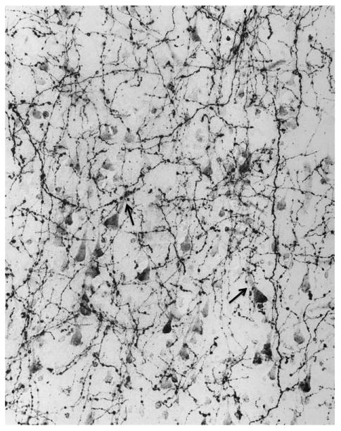
This photomicrograph of AChE histochemistry based on a modified Karnovsky-Roots method shows cholinergic axons in layer 3 of inferotemporal cortex in the brain of a 22-year-old control specimen. The multiple varicosities represent putative sites of ACh release and are closely associated with AChE-rich cholinoceptive pyramidal neurons. The arrows point to two examples where the varicosities are apposed to the apical dendrite of cholinoceptive neurons. ×415. From Mesulam and Geula (1988) with permission.