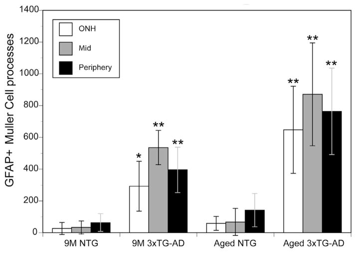
Fig. 2. The number of GFAP-positive Müller cell processes was significantly increased in the 3xTG-AD retina compared to the NTG retina.
The average number of GFAP-positive Müller cell processes in a 20× field was counted at the optic nerve head (ONH), mid, and peripheral retina of 9 M and aged (18–24 M) 3xTG-AD and NTG mice. There was a significant increase (*indicates p < 0.01, ** indicates p ≤ 0.001; n = 3 per region per group) in all regions of the retina at both time points investigated. Also notable is the slight but non-significant increase in GFAP-positive Müller cells with age observed in the NTG mice. This effect was more drastic, but still not significant, with age in the 3xTG-AD mice.

