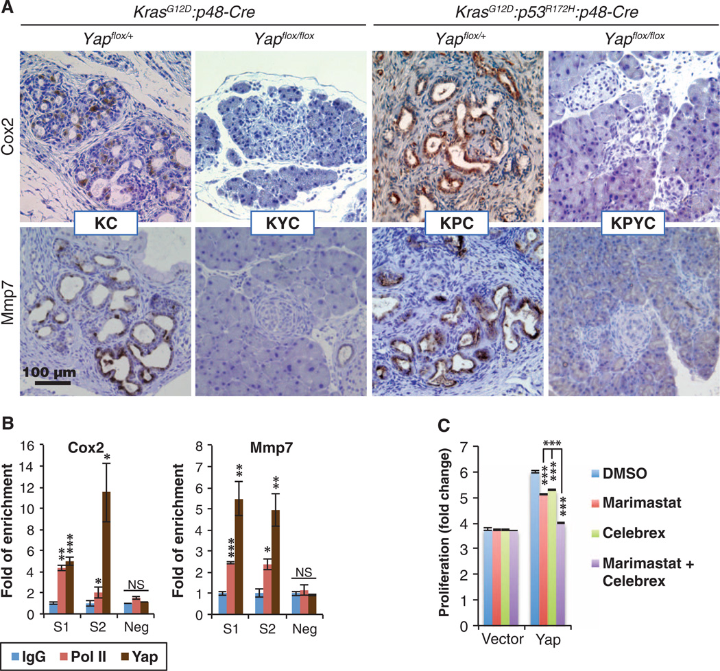
Fig. 5. Yap controls the expression of Cox2 and Mmp7 in vitro and in vivo.
(A) Representative images of IHC staining for Cox2 and Mmp7 in pancreatic sections from KC, KPC, KYC, and KPYC mice. Scale bar, 100 µm. (B) qRT-PCR analysis of ChIP with antibodies to immunoglobulin G (IgG), polymerase II (Pol II), and Yap on Cox2 and Mmp7 promoter regions that contain (S1 and S2) or do not contain (neg) putative TEAD-binding sites in Yap-reconstituted KPY cells. Data are means ± SD from three independent experiments. (C) Fold proliferation in KPY cells expressing control or Yap vector 3 days after addition of DMSO (control), marimastat (MMP inhibitor, 5 µM), or Celebrex (Cox2 inhibitor, 10 µM). Data are means ± SD from three independent experiments. *P< 0.01, **P< 0.001, ***P< 0.0001, two-tailed t test.

