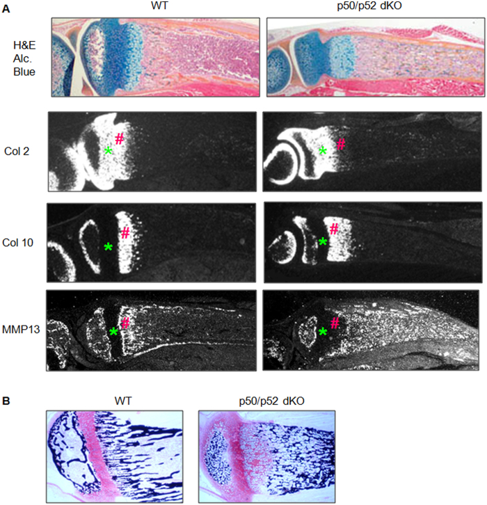
Figure 2.
Expression of chondrocyte marker genes in tibial growth plates from 2-week-old NF-kB p50/p52 dKO and littermate control mice. (A) In-situ hybridization showing the distribution of type-2 and −10 collagen (Col), and MMP13 mRNA. The proliferative chondrocyte zone is indicated by * and the hypertrophic chondrocyte zone is indicated by #. (B) Safranin O/von Kossa-stained plastic-embedded sections showing a thickened hypertrophic chondrocyte zone and increased volume of mineralized bone matrix in p50/p52 dKO mice.

