
Figure 4.
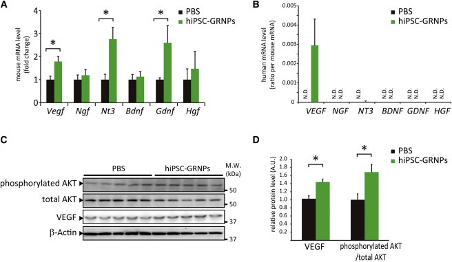
hiPSC-GRNPs Transplantation Increased Neurotrophic Factors and Activated AKT Signal
(A) Gene expressions of neurotrophic factors, including Vegf, Ngf, Nt3, Bdnf, Gdnf, and Hgf were quantitatively analyzed using mouse-specific primers. The levels of Vegf, Nt3, and Gdnf were significantly increased in the hiPSC-GRNPs transplantation group (∗p < 0.05). Data represent mean ± SD (n = 3 mice per group).
(B) Gene expressions of neurotrophic factors, including VEGF, NGF, NT3, BDNF, GDNF, and HGF were quantitatively analyzed using human-specific primers and the human-origin/mouse-origin ratio was calculated. Data represent mean ± SD (n = 3 mice per group).
(C) Western blot analysis of phosphorylated AKT, total AKT level, and VEGF in lumbar spinal cord in the PBS injection group and hiPSC-GRNPs transplantation group.
(D) Densitometric analysis of (C). Measured values of proteins were normalized by that of β-actin. The levels of VEGF and AKT phosphorylation were significantly increased in the hiPSC-GRNPs transplantation group (∗p < 0.05).
Data represent mean ± SD (n = 5 mice per group). N.D., not detectable. See also Figure S3 and Table S2.