Figure 3.

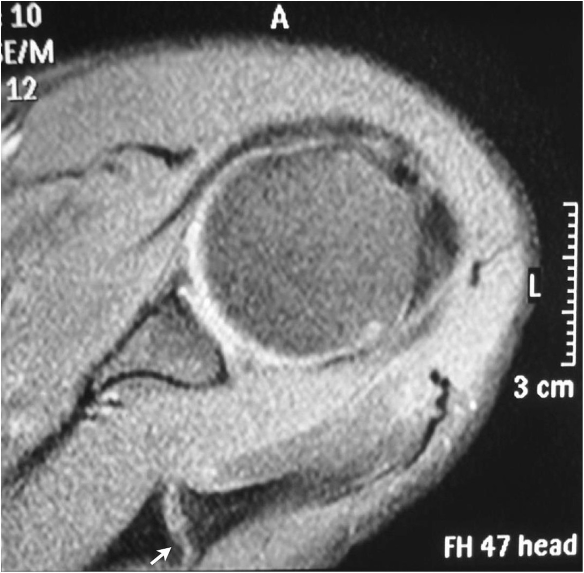
Preoperative magnetic resonance imaging (MRI). An axial section of the T2-weighted MRI demonstrating a hypertrophic pseudarthrosis of the fracture of the base of the acromion (arrow).

Preoperative magnetic resonance imaging (MRI). An axial section of the T2-weighted MRI demonstrating a hypertrophic pseudarthrosis of the fracture of the base of the acromion (arrow).