
Figure 8.
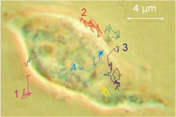
Trajectories of single AAV-Cy5 particles indicating infectious entry pathways of AAVs into a living cervical cancer cell line (HeLa). The traces showing single diffusing virus particles were recorded at different times. They describe various stages of AAV infection, e.g. diffusion in solution (1 and 2), touching at the cell membrane (2), penetration of the cell membrane (3), diffusion in the cytoplasm (3 and 4), penetration of the nuclear envelope (4), and diffusion in the nucleoplasm. Reprinted with permission from AAAS.[111]