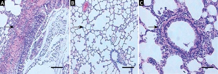
Figure 1.
Histopathology of lung tissue of different groups of rats. A – In AP rats, the lung tissue showed a significantly wider alveolar septum, and a large number of infiltrated white blood cells. B – In control rats, the lung tissue was normal. C – After PAG treatment, the pulmonary interstitial edema, alveolar exudation, and bleeding were significantly decreased. Arrows indicate white blood cells. Bar: 100 μM

