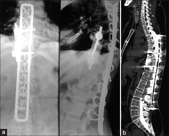
Figure 1.

(a) Plain radiographs anteroposterior and lateral views showing instrumentation with Hartshill and sublaminar wires showing fusion at D10-D11 and no implant breakage or cut-out (b) Computed tomography myelogram showing complete cut off of dye flow at L3-L4 level
