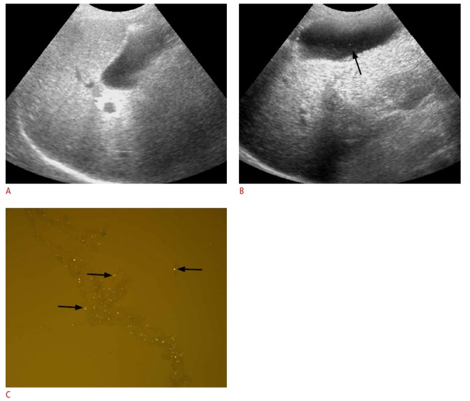
Fig. 5. A 76-year-old woman with acute cholecystitis.

A. Fundamental ultrasonography shows no stones in the gallbladder lumen. B. Harmonic ultrasonography with a high background noise shows microlithiasis (arrow) in the gallbladder lumen. C. Gallbladder fluid was percutaneously aspirated and centrifuged. The precipitates show clear microcrystals with some colloid components (arrows) on polarized microscopy (×200).
