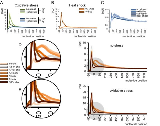
Figure 2.
Ribosomal occupancy profiles and the effect of stress and drug treatment. (A) Control yeast cells and cells treated with hydrogen peroxide (0.2 mM) in the presence or absence of 100 μg/ml cycloheximide in the media. Nucleotide position count is relative to start codon. (B) Ribosome occupancy profiles of yeast cells undergoing heat shock (42°C, 20 min). The peak appears only when cycloheximide is added to the medium. (C) None of the three tested stress types lead to a significant increase of ribosomes at the 5′ proximities of reading frames in the absence of cycloheximide. Refer to Figure 3A and B for additional details on amino acid starvation. (D and E) Concentration of cycloheximide in the medium affects the shape of the profile, pointing to a passive diffusion model of cycloheximide entering live cells. Cells were grown in YPD medium in the absence of stress (D) or subjected to oxidative stress (0.2 mM hydrogen peroxide, 30 min) (E). Cycloheximide concentration does not immediately reach the threshold, under which all ribosomes are inhibited with 100% efficiency, instead increasing gradually. Therefore, following the treatment some ribosomes initiating translation continue protein synthesis until they encounter the drug, leading to a broad cumulative peak in the ribosomal occupancy profile.

