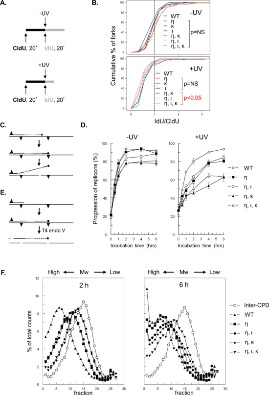
Figure 1.

Both Pols ι and κ are required for replicon progression in Polη-deficient MEFs, late after UVC exposure. (A) Schematic representation of DNA fiber labeling with nucleotide analogs CldU and IdU in MEFs that were mock treated (-UV) or exposed to UVC (+UV). (B) Cumulative percentage of replication forks at ratios of lengths of ldU-labeled tracts to CldU-labeled tracts in wild-type MEFs (WT) or in MEFs with single, double or triple deficiencies in Polη (η), Polι (ι) and Polκ (κ) exposed to 13 J/m2 UVC (+UV). P values are shown of the two-sample Kolmogorov-Smirnov (K-S) test for the ratio distribution of each knock-out genotype compared to wild-type. (C) Scheme of the alkaline DNA unwinding assay. Nascent DNA is pulse labeled with [3H]thymidine (dotted line) immediately before the induction of photolesions (triangles; top). MEFs are then cultured in medium without label (middle). Stalling of a fork at a photolesion results in a DNA end containing [3H]thymidine that is locally denatured using alkaline, followed by sonication and isolation of [3H]thymidine-labeled ssDNA using hydroxyl apatite (bottom). (D) Replication fork progression in mock-treated MEFs (left panel) and in MEFs exposed to 5 J/m2 UVC (right panel; n = 4). Error bar, SEM. (E) Scheme of alkaline sucrose gradient sedimentation using T4 endonuclease V. Template DNA was uniformly labeled with [14C]thymidine (solid line) followed by exposure to UVC inducing CPD and (6–4)PP photolesions (triangles; top). Elongating daughter strands were pulse labeled with [3H]-thymidine for 30 min (dotted line) and cultured in a medium without label (dashed line; middle). At different times, cells were lysed and [14C]thymidine-containing DNA was cleaved by T4 endonuclease V at a CPD, followed by size fractionation using alkaline sucrose gradients (bottom). The [14C]thymidine-labeled inter-CPD size distribution serves as an internal standard, since CPDs are not removed in mouse cells. (F) Alkaline sucrose gradient profiles of [3H]thymidine-containing DNA of wild-type MEFs (WT, closed triangle), MEFs deficient in Polη (η; closed square) and of Polη-deficient MEFs containing an additional defect in Polι (ι, η; closed circle), Polκ (η, κ; closed diamond) or both TLS polymerases (η, ι, κ; closed inverted triangle) at 2 and 6 h after exposure to 5 J/m2 UVC. Also the profile of [14C]thymidine labeled, CPD-containing fragments is depicted (open circles). Mw, molecular weight.
