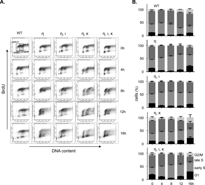
Figure 2.

The replicative activity of Polη-deficient MEFs exposed to UVC relies mainly on Polκ. (A) FACS profiles showing BrdU content of wild-type MEFs (WT), MEFs deficient in Polη (η) and Polη-deficient MEFs containing an additional defect in Polι (η, ι), Polκ (η, κ) or both TLS polymerases (η, ι, κ) after exposure to 5 J/m2 UVC. Prior to fixation, MEFs were pulse labeled with BrdU for 30 min, immediately or at 4, 8, 12 and 16 h after UVC treatment. BrdU incorporation was determined by immunostaining and DNA content was measured using propidium iodide. (B) Quantification of MEFs in different cell cycle stages, up to 16 h after UVC exposure (n = 3; error bars: SD).
