
Figure 3.
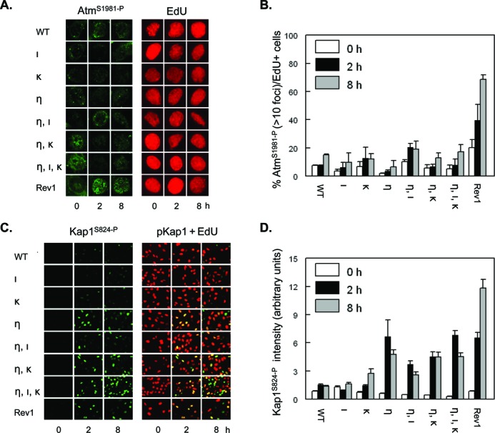
Few UVC-induced dsDNA breaks in Polη-deficient MEFs with or without additional deficiencies in Pols ι and κ. (A) Wild-type MEFs (WT), MEFs with single, double or triple deficiencies in Polη (η), Polι (ι) and Polκ (κ), or MEFs deficient in Rev1 (Rev1) were pulse labeled with EdU for 30 min, prior to exposure to 5 J/m2 UVC. Then, MEFs were fixed at 0, 2 and 8 h after treatment and immunostained for AtmS1981-P (left panel, online in green) in replicating, EdU-incorporating MEFs (online in red) at the time of UVC exposure. (B) Quantification of EdU-positive MEFs containing at least 10 AtmS1981-P foci. Error bar, SEM. (C) Similar experiment as in (A), except that MEFs were immunostained for Kap1S821-P (left panels, online in green) in replicating, EdU-incorporating MEFs (right panels, merge of staining for Kap1S821-P (online in green) + EdU (online in red)). (D) Quantification of the intensity of Kap1S821-P signal in EdU-positive MEFs. Error bars, SEM.