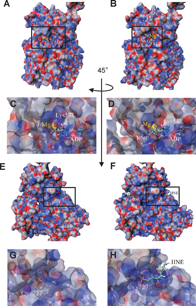
Figure 8.

Changes in molecular surface of helicase and RQC domains of WRN protein upon HNE adduction. Surface representation of WRN electrostatic potential (±25 kT/e, red-negative, blue-positive, gray-neutral) before (A) and after HNE addition to Lys577 (B). A comparison of the vicinity of native (C) and HNE adducted Lys577 (D) show some changes in molecular surface in the proximity of the ATP (ADP) binding pocket. (E) Surface representation of helicase and RQC domains of the WRN protein after orthogonal rotation along the Y-axis of 45° in relation to structure shown in (A). (F) Surface representation of helicase and RQC domains of the WRN protein with HNE attached to Cys727. Detailed views of the vicinity of native (G) and modified (H) Lys727 illustrate changes in charge distribution and reveal surface location of HNE.
