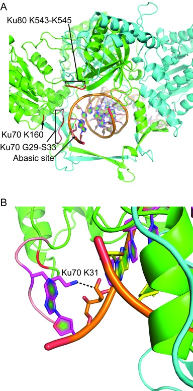
Figure 6.

Model of the interaction between Ku70 K31 and an abasic site substrate. A and B. A cartoon representation of the human Ku heterodimer interacting with an abasic site substrate was modeled on the structure 1JEY (21). Five amino acids (pink) were appended to the most N-terminal residue resolved in Ku70 (G34), and the DNA altered to have an abasic site centered within a three nucleotide 5′ overhang. A. Ku70 is in green and Ku80 in cyan. The location of candidate nucleophiles identified in Figures 1 and 2 are shown in red. B. Ku70 K31 is shown in stick representation, with the dashed line showing the distance between nuclei predicted to participate in a Schiff base intermediate.
