
Figure 4.
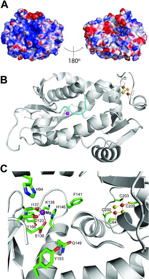
Structure of the active site of Pcal_0546. (A) Surface charge distribution of Pcal_0546 with a blue (positively charged) to red (negatively charged) gradient. (B) The orientation of the loop (138–142) located at the bottom of the active site of Pcal_0546 (as opposed to the open active site in SSO0001). The Mg2+ ion is represented by the magenta sphere. (C) Close-up view of the Pcal_0546 active site. Residues are shown as sticks and labeled, the Mg2+ ion is shown as a purple sphere, and the [2Fe-2S] cluster as orange and yellow spheres.