
Figure 2.
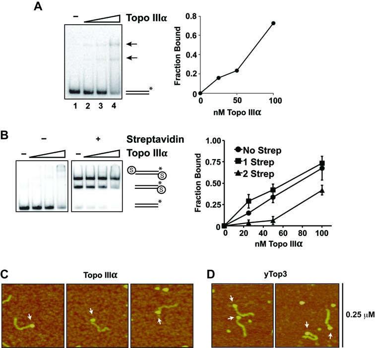
Recognition of DNA ends by Topo IIIα. (A) Topo IIIα (25, 50 or 100 nM) was incubated with radiolabeled 80-mer dsDNA (5 nM ends). Quantification of DNA binding is shown in the right panel. The arrow denotes nucleoprotein complexes and the asterisk indicated the location of the radiolabel in the DNA substrate. (B) DNA mobility shift was conducted as in (A), except that dsDNA substrate blocked by biotin-streptavidin (denoted by the circled S) at either one or both of the ends was used. The data shown were the average from three independent experiments and the error bars represent 1 SD. (C, D) AFM was used to image nucleoprotein complexes of Topo IIIα and Top3. Arrows indicate DNA end binding events.