
Figure 1.
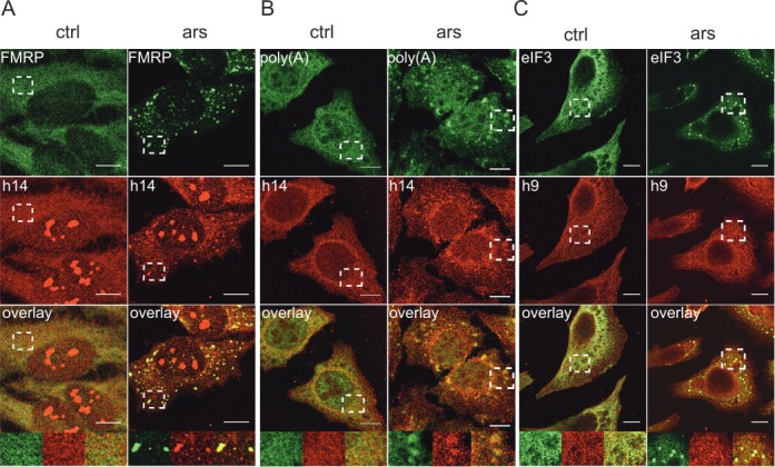
SGs contain SRP9/14. Immunofluorescence images of HeLa cells stained with antibodies against human SRP14 (h14), SRP9 (h9) and against different SG markers. ars: cells treated with 500 μM sodium arsenite for 30 min; ctrl: untreated cells. (A) Antibodies against h14 and FMRP. (B) Anti-h14 antibodies and in situ hybridization of mRNAs with oligo(dT). (C) Anti-h9 and anti-eIF3 antibodies. Anti-h9 antibodies failed to stain nucleoli. (A–C) Images were captured at the SP2 laser scanning confocal microscope (63x/1.4 numerical aperture, PlanApo). Areas denoted by rectangles are shown at higher magnification. Scale bars: 10 μm.