
Figure 2.
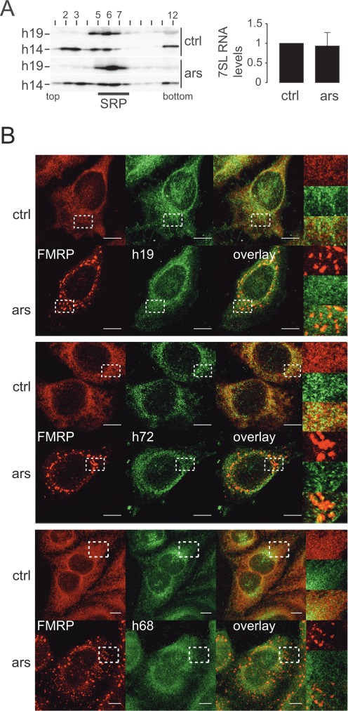
The SRP complex remains intact during stress and does not localize to SGs. (A) Velocity sedimentation fractionations of HeLa cell postnuclear supernatants on 12–30% glycerol gradients. Left panel: Western blots of gradient fractions using anti-h14 and anti-h19 antibodies. Right panel: quantification of 7SL RNA in HeLa cell extracts by qRT-PCR. Levels were standardized to GAPDH mRNA and normalized to control cells. Error bars are shown as SD, n = 3. (B) Immunofluorescence images of HeLa cells stained with antibodies against h19 and FMRP (upper panel) against h72 and FMRP (middle panel) and against h68 and FMRP (lower panel). Images were captured using a 63x lens on LSM-710 Laser scanning microscope. Areas denoted by rectangles are shown at higher magnification. Scale bars: 10 μm. ars: 500 μM sodium arsenite for 30 min; ctrl: untreated cells. h19: human SRP19; h72: human SRP72; h68: human SRP68