Figure 4.

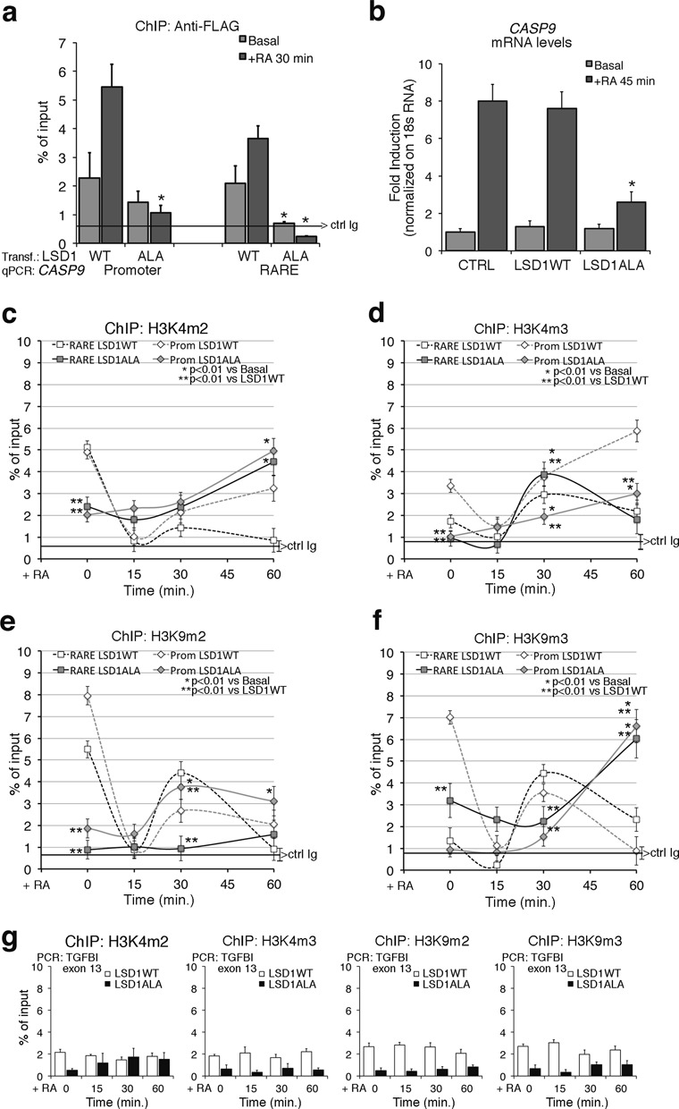
Expression of a dominant negative LSD1 mutant inhibits RA-induced methylation changes and CASP9 induction by RA. (a) Recruitment of wild type (WT) and mutant LSD1 (LSD1ALA) to the CASP9 RARE–promoter. The LSD1ALA mutant contains a substitution of threonine with alanine in position 110 and has been described elsewhere (17,19). MCF7 were transiently transfected with LSD1 vectors (WT or mutant), starved (Basal) or treated with 300-nM RA for 30 min and were analyzed by qChIP using Anti-FLAG antibodies to recognize the recombinant LSD1. The panel shows the recruitment of the flagged LSD1WT and LSD1ALA to the RARE and to the promoter sequences of CASP9. The black, horizontal line indicates the percent of input from a control ChIP (Ab: non-immune serum). (b) LSD1ALA inhibits RA-induced CASP9 transcription. Total RNA was prepared from MCF7 transiently transfected with LSD1WT or LSD1ALA. After 48 h, mRNAs from control cells (starved) or RA-induced cells (300-nM RA for 45 min) were analyzed by qPCR with specific primers to CASP9 mRNA. (c, d) LSD1ALA inhibits RA-induced H3K4 demethylation. H3K4me2 and H3K4me3 occupancy at the promoter and RARE of CASP9 in LSD1ALA expressing cells as in (b). (e, f) LSD1ALA inhibits RA-induced H3K9 demethylation. H3K9me2 and H3K9me3 occupancy at the promoter and RARE of CASP9 in LSD1ALA expressing cells as in (b). (g) ChIP analysis of TGFBI exon 13 (non-RA-induced gene), in transfected cells exposed to RA for 15, 30 and 60 min. For each antibody, the control IgG signal is shown (brackets ± SD). The statistical analysis derives from at least three experiments in triplicate (n ≥ 9; mean ± SD); *P < 0.01 (matched pairs t-test): compared to the RA-unstimulated sample; **P < 0.01 (student t-test): comparison between control and the LSD1ALA expressing cells at the same time.
