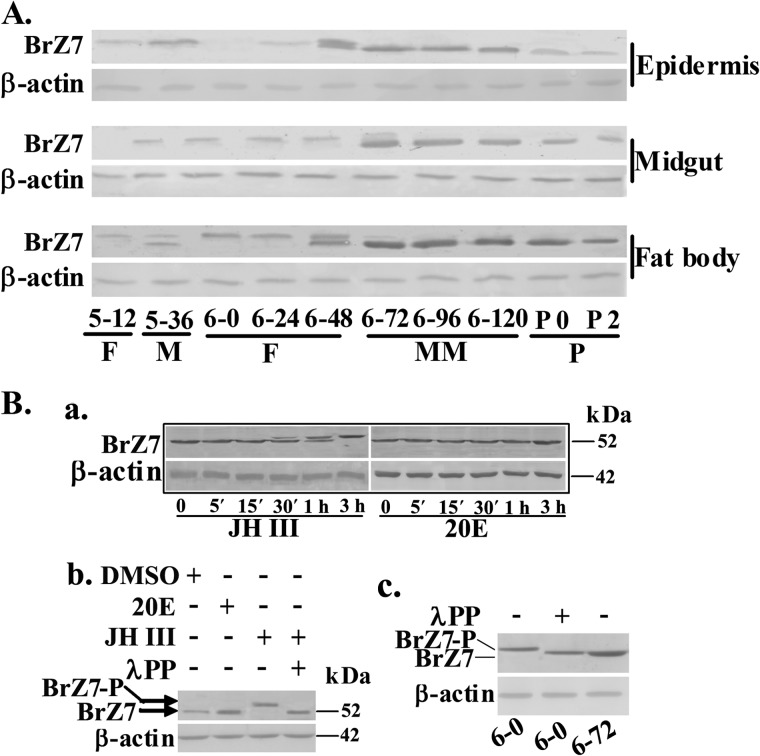
FIGURE 4.
JH-induced BrZ7 phosphorylation analyzed by Western blot. The SDS-PAGE gel was 7.5%. A, the expression profiles of BrZ7 during development were detected using anti-BrZ7 antibody. 5–12 to 6–120 indicate the larval developmental stages from fifth instar 12 h to 6th-120 h, respectively. P0–P2, days of pupal development. F, feeding; M, molting; MM, metamorphic molting; P, pupae. B, a, cells were incubated with 1 μm 20E or JH III for 5, 15, 30, 60, and 180 min, respectively. Proteins were then extracted for Western blot analysis with anti-BrZ7 antibody. b, cells were treated with 1 μm 20E or JH III for 3 h; proteins were isolated and incubated with λ-phosphatase at 5 μm for 30 min. In c, 6–0 indicates the proteins from the fat body of 6th instar 0 h larvae, and 6–72 indicates the proteins from 6th instar 72 h larvae, analyzed by Western blot. BrZ7-P, phosphorylated BrZ7.

