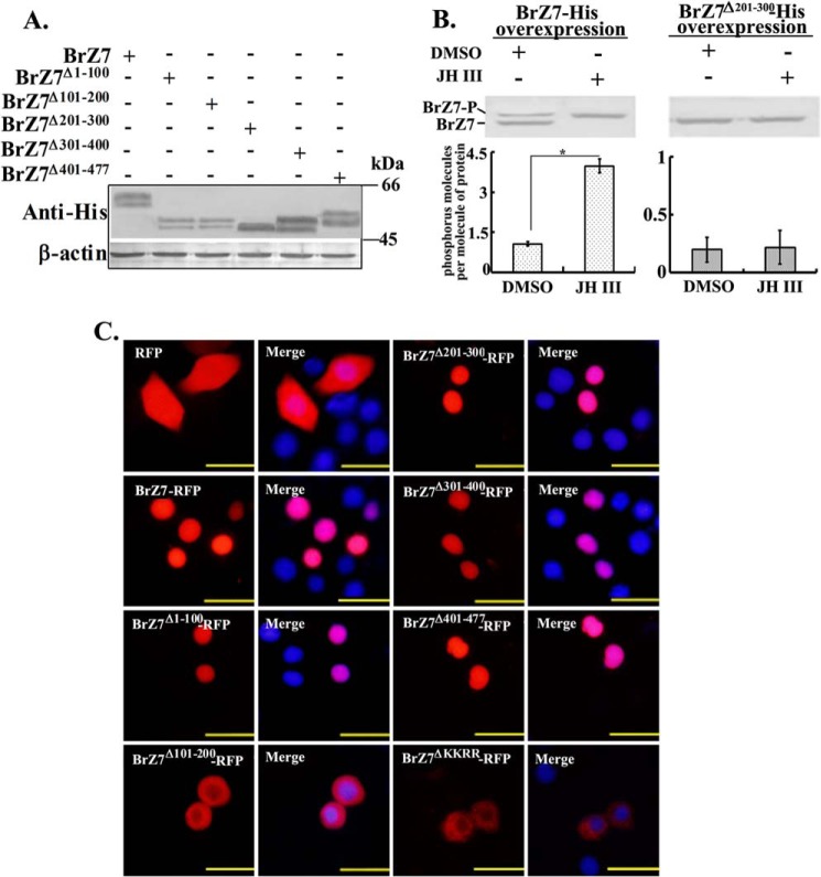
FIGURE 6.
Analysis of the phosphorylation sites and subcellular location of BrZ7. A, cells were transfected with various plasmids expressing different BrZ7 mutants fused with His tag. Proteins were extracted, and Western blot was performed with anti-His antibody using 7.5% SDS-polyacrylamide gel. B, number of molecules of phosphorus per molecule of BrZ7-His or BrZ7Δ201–300-His by DMSO or JH III treatments analyzed by a phosphoprotein phosphate estimation assay kit. The cells were transfected with pIEx-4-BrZ7-His or pIEx-4-BrZ7Δ201–300-His plasmids in HaEpi cells. After 48 h of overexpression, the cells were incubated with DMSO or 1 μm JH III for 3 h, and the proteins were purified by His-Bind resin for the measurement. About 50 μg of protein was used per assay. Statistical significance (*, p < 0.05) was based on three biologically independent repeats and analyzed by the Student's t test. C, BrZ7 and various truncated BrZ7-RFP mutants were overexpressed in HaEpi cells, and pIEx-4-RFP-transfected plasmid was used as a control sample. The nuclei were stained with DAPI. Fluorescence signal was visualized using an Olympus BX51 fluorescence microscope. Scale bar, 25 μm. Error bars, S.E.

