FIGURE 6.
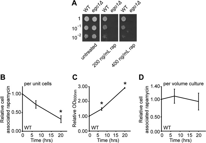
Rapamycin is stable in wild-type cells and decreases only with residual proliferation during recovery. Cultures of WT and ego1Δ cells in mid-logarithmic phase were diluted to an A600 nm of ∼0.1 and treated or not for 4 h with rapamycin (rap) (either 200 or 400 ng/ml) at room temperature. Cells were washed in YPD and serial dilutions were spotted (5 μl) onto YPD plates and incubated at 30 °C for 2 days. Scans of the plates are shown in A. Exponentially growing cultures of WT cells in YPD at an A600 nm of 0.6 were treated with rapamycin (400 ng/ml) for 4 h at room temperature after which cells were washed three times and transferred into fresh YPD to recover. Samples were taken at 0, 7, and 20 h following drug removal and diluted as appropriate to prevent saturation. B, relative cell-associated signal for rapamycin in WT cells as a function of recovery time and expressed relative to the mass spectrometry signal at t = 0. C, relative increase in culture density (by A600 nm) for WT cells as a function of recovery time. D, relative cell-associated rapamycin signal for WT cells as a function of recovery time (from B) and corrected for growth of the culture (from C). Error bars denote S.E.; *, p < 0.05 relative to t = 0.

