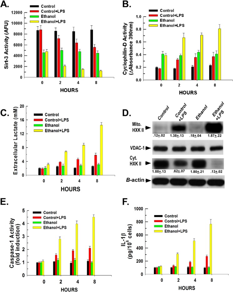
FIGURE 2.
LPS and ethanol enhance hexokinase II binding to the mitochondria in Kupffer cells by modulating the activity of sirtuin-3 and cyclophilin-D. A, Kupffer cells were plated in 6-well plates at 500,000 cells/well. Following a 16-h incubation, the cells were either left untreated or treated with 100 ng/ml LPS. At the time points indicated, the cells were harvested, and mitochondria were isolated. Sirtuin-3 activity was determined in mitochondrial extracts as described under “Experimental Procedures.” Values are the means of three independent experiments with the error bars indicating S.D. B, Kupffer cells were plated in 6-well plates at 500,000 cells/well. Following a 16-h incubation, the cells were left untreated or treated with 100 ng/ml LPS. At the time points indicated, the cells were harvested, and mitochondria were isolated. Mitochondrial extracts were prepared, and cyclophilin-D activity was determined as described under “Experimental Procedures.” C, Kupffer cells were plated in 24-well plates at 50,000 cells/well. After 16 h, the cells were either left untreated or treated with 100 ng/ml LPS. At the time points indicated, aliquots of medium were collected, and the concentration of lactate was determined colorimetrically as described under “Experimental Procedures.” Values are the means of three independent experiments with the error bars indicating S.D. D, Kupffer cells were plated in 6-well plates at 500,000 cells/well. After 16 h, the cells were either left untreated or treated with 100 ng/ml LPS. After 1 h, the cells were harvested, and cytosolic (Cyt.) and mitochondrial (Mito.) fractions were prepared and used for Western blotting. Densitometry values are indicated below their respective bands and are the means of three independent experiments ±S.D. E, Kupffer cells were plated in 24-well plates at 50,000 cells/well. Following 16 h of incubation, the cells were either left untreated or treated with 100 ng/ml LPS. At the time points indicated, the cells were harvested, and caspase-1 activity was determined fluorescently as described under “Experimental Procedures.” Values are the means of three independent experiments with the error bars indicating S.D. F, Kupffer cells were plated in 24-well plates at 50,000 cells/well. Following 16 h of incubation, the cells were either left untreated or treated with 100 ng/ml LPS. At the time points indicated, medium aliquots were taken and assessed for IL-1β levels by ELISA. Values are the mean of three independent experiments with the error bars indicating S.D. AFU, arbitrary fluorescence units; VDAC-1, voltage-dependent anion channel 1.

