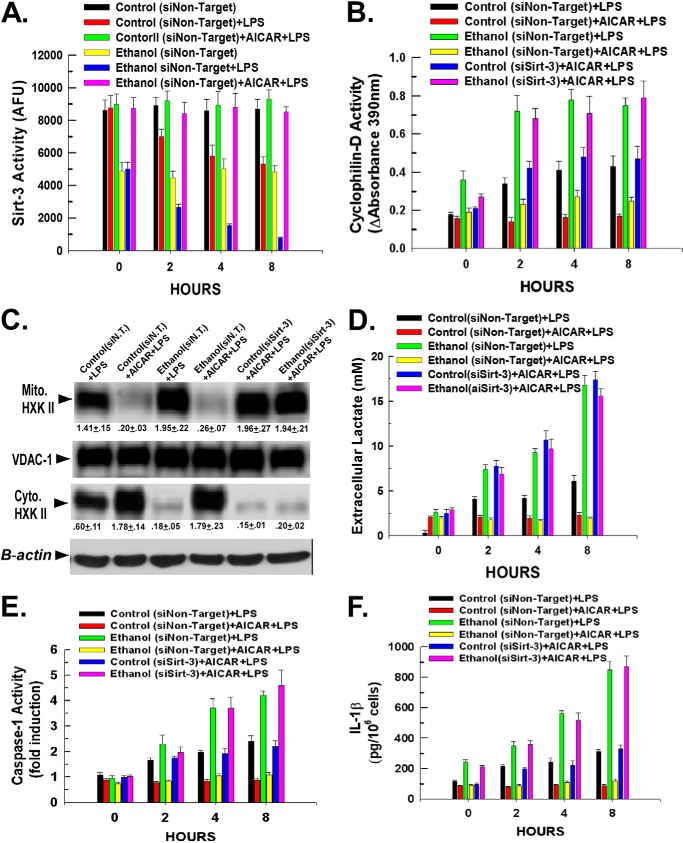
FIGURE 6.
Activation of AMPK reverses the inhibition of sirt-3 activity mediated by LPS and ethanol and the potentiating effect on Kupffer cell stimulation. A, Kupffer cells isolated from control- or ethanol-fed rats were plated in 6-well plates at 500,000 cells/well and incubated for 16 h. Following incubation, the cells were transfected with 50 nm non-targeting siRNA. After 24 h, the cells were then treated with 100 ng/ml LPS. Alternatively, the cells were pretreated with 0.5 mm AICAR for 30 min prior to exposure to LPS. At the time points indicated, the cells were harvested, and mitochondria were isolated. Mitochondrial lysates were prepared, and sirtuin-3 activity was determined as described under “Experimental Procedures.” Values are the means of three independent experiments with the error bars indicating S.D. B, Kupffer cells isolated from control- or ethanol-fed rats were plated in 6-well plates at 500,000 cells/well and incubated for 16 h. The cells were then transfected with 50 nm non-targeting siRNA or siRNA targeting sirtuin-3 and incubated for a further 24 h. The cells were then treated with 100 ng/ml LPS. Alternatively, the cells were pretreated with 0.5 mm AICAR for 30 min prior to exposure to LPS. At the time points indicated, the cells were harvested, and mitochondria were isolated. Mitochondrial lysates were prepared, and cyclophilin-D activity was determined as described under “Experimental Procedures.” Values are the means of three independent experiments with the error bars indicating S.D. C, Kupffer cells isolated from control- or ethanol-fed rats were plated in 6-well plates at 500,000 cells/well and incubated for 16 h. The cells were then transfected with 50 nm non-targeting siRNA (siN.T.) or siRNA targeting sirtuin-3 and incubated for a further 24 h. The cells were then treated with 100 ng/ml LPS. Alternatively, the cells were pretreated with 0.5 mm AICAR for 30 min prior to exposure to LPS. After 1 h, the cells were harvested, and mitochondria were isolated. Mitochondrial (Mito.) and cytosolic (Cyt.) lysates were prepared and utilized for Western blotting. Densitometry values are indicated below their respective bands and are the mean of three independent experiments ±S.D. D, Kupffer cells isolated from control- or ethanol-fed rats were plated in 24-well plates at 50,000 cells/well and incubated for 16 h. The cells were then transfected with 50 nm non-targeting siRNA or siRNA targeting sirtuin-3 and incubated for a further 24 h. The cells were then treated with 100 ng/ml LPS. Alternatively, the cells were pretreated with 0.5 mm AICAR for 30 min prior to exposure to LPS. At the time points indicated, aliquots of medium were taken, and the concentration of lactate was determined colorimetrically as described under “Experimental Procedures.” Values are the means of three independent experiments with the error bars indicating S.D. E, Kupffer cells isolated from control- or ethanol-fed rats were plated in 24-well plates at 50,000 cells/well and incubated for 16 h. The cells were then transfected with 50 nm non-targeting siRNA or siRNA targeting sirtuin-3 and incubated for a further 24 h. The cells were then treated with 100 ng/ml LPS. Alternatively, the cells were pretreated with 0.5 mm AICAR for 30 min prior to exposure to LPS. At the time points indicated, the cells were harvested, and caspase-1 activity was determined fluorescently in whole cell lysates. Values are the means of three independent experiments with the error bars indicating S.D. F, Kupffer cells isolated from control- or ethanol-fed rats were plated in 24-well plates at 50,000 cells/well and incubated for 16 h. The cells were then transfected with 50 nm non-targeting siRNA or siRNA targeting sirtuin-3 and incubated for a further 24 h. The cells were then treated with 100 ng/ml LPS. Alternatively, the cells were pretreated with 0.5 mm AICAR for 30 min prior to exposure to LPS. At the time points indicated, the cells were harvested, and caspase-1 activity was determined fluorescently in whole cell lysates by ELISA as described under “Experimental Procedures.” Values are the means of three independent experiments with the error bars indicating S.D. AFU, arbitrary fluorescence units; VDAC-1, voltage-dependent anion channel 1.

