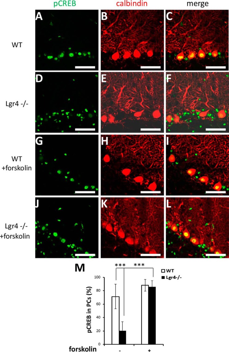
FIGURE 9.
Decreased activation of Creb in PCs of Lgr4−/− mice. Double immunofluorescence staining for anti-phosphorylated Creb antibody (green) and anti-calbindin (red) antibody in the cerebellar slices of wild-type (A–C) and Lgr4−/− (D–F) mice. Wild-type (G–I) and Lgr4−/− (J–L) slices were perfused for 10 min with 50 μm forskolin. Similar staining patterns were observed throughout the cerebellar cortices of wild-type and Lgr4−/− mice. M, statistical analysis showed that pCreb in PCs was selectively decreased in Lgr4−/− mice. The statistical data were obtained from five adjacent midsagittal sections/mouse and five mice/genotype. Data are mean ± S.E., Student's t test was used for statistical analysis, ***, p < 0.001. Scale bar, 50 μm.

