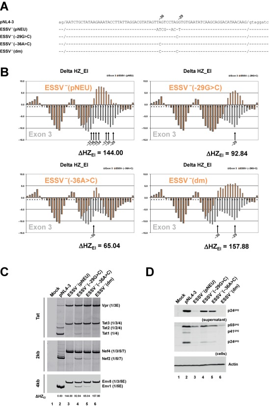
Figure 6.

HEXplorer screening identifies minimal point mutations disrupting ESSV. (A) pNL4-3-derived wild-type and mutated HIV-1 exon 3 sequences. Nucleotide coordinates refer to the 3’ end of exon 3. (dm) refers to the double mutation −29G>C and -36A>C. (B) HZEI score profiles of HIV exon 3 reference pNL4–3 (gray) versus mutant sequences ESSV− (pNEU), ESSV− −29G>C, ESSV− −36A>C and ESSV− (dm) (red). HEXplorer score differences are given below the profile graphs, and point mutations are indicated by black arrows and corresponding positions. (C) RT-PCR analysis of splicing patterns for different classes of viral RNAs. 2.5 × 10⁵ HEK 293T cells were transiently transfected with 1 μg of each of the proviral DNAs. Thirty hours after transfection, total RNA was isolated from the cells and subjected to RT-PCR analyses with different sets of primer pairs covering intronless and intron-containing mRNA classes described elsewhere (20). RT-PCR products were separated by 8% non-denaturing polyacrylamide gel electrophoresis and visualized using ethidium bromide staining. HIV-1 mRNA isoforms are indicated on the right and correspond to the nomenclature published previously (41). HZEI-score differences (ΔHZEI) with respect to the reference pNL4-3 are given below the gel. (D) Western blot analysis of Gag expressed by reference and mutant proviruses. 2.5 × 10⁵ HEK 293T cells were transiently transfected with 1 μg of each of the proviral DNAs. Forty-eight hours post transfection viral supernatants were collected, layered onto 20% sucrose solution and centrifuged at 28 000 rpm for 1.30 h at 4°C to pellet the released viral particles. In addition, cells were harvested and resuspended in lysis buffer. Supernatants and cellular lysates were resolved in 12% SDS-PAGE and electroblotted on nitrocellulose membranes. To determine virus particle production and the expression of viral proteins, samples were probed with a primary antibody against HIV-1 p24 (Biochrom AG). Equal amounts of cell lysates were controlled by the detection of α-actin (Sigma-Aldrich, A2228).
